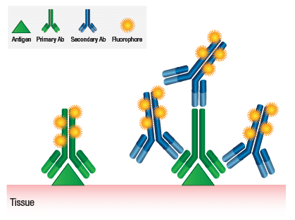
Immunohistochemistry (IHC) staining with fluorescent antibodies, also called immunofluorescence (IF), visualizes specific proteins or antigens in tissues by using antibodies tagged with fluorophores (fluorescent dyes). In a dissected Drosophila brain—a small (~200–300 µm), whole-mount tissue—this technique reveals neural architecture, gene expression, or protein localization with high resolution, often via confocal microscopy.
The process begins with dissection. Adult fly heads are removed under a stereomicroscope in cold phosphate-buffered saline (PBS) or artificial cerebrospinal fluid. The brain is gently extracted from the cuticle, avoiding damage, and trachea removed to prevent floating or imaging artifacts.
Next, fixation preserves structure and antigens. Brains are typically immersed in 4% paraformaldehyde (PFA) in PBS or PBT (PBS + 0.3% Triton X-100) for 30–60 minutes on ice or at room temperature. Triton X-100 permeabilizes membranes for antibody penetration.
After washing in PBT (multiple 5–10 min changes), blocking reduces non-specific binding. Samples incubate in 5% normal goat serum (NGS) or similar in PBT for 1–2 hours.
Primary antibody incubation targets the antigen of interest (e.g., anti-GFP for transgenic reporters, anti-Brp for synapses, or anti-nc82 for neuropil). Diluted in blocking buffer, brains incubate overnight (or longer) at 4°C on a nutator for gentle agitation.
Washes (5–6× in PBT, 10–30 min each) remove unbound primary antibody.
Secondary antibody—conjugated to fluorophores like Alexa Fluor 488 (green), 555 (red), or 647 (far-red)—binds the primary antibody’s species (e.g., anti-rabbit Alexa 488). Incubation lasts 1–2 days at 4°C. Multiple secondaries enable multi-color labeling (e.g., one channel for neurons, another for glia).
Final washes remove excess secondary antibody. Optional counterstains like DAPI label nuclei (blue).
Brains mount between coverslips in anti-fade media (e.g., Vectashield) for imaging. Confocal or light-sheet microscopy captures z-stacks, producing 3D reconstructions of fluorescent signals.
This method’s strength lies in specificity and multiplexing—e.g., labeling neurotransmitters, synaptic markers, or mutant phenotypes in the compact fly brain. Challenges include antibody penetration in dense tissue and background fluorescence, mitigated by optimized permeabilization and controls.

Leave a Reply