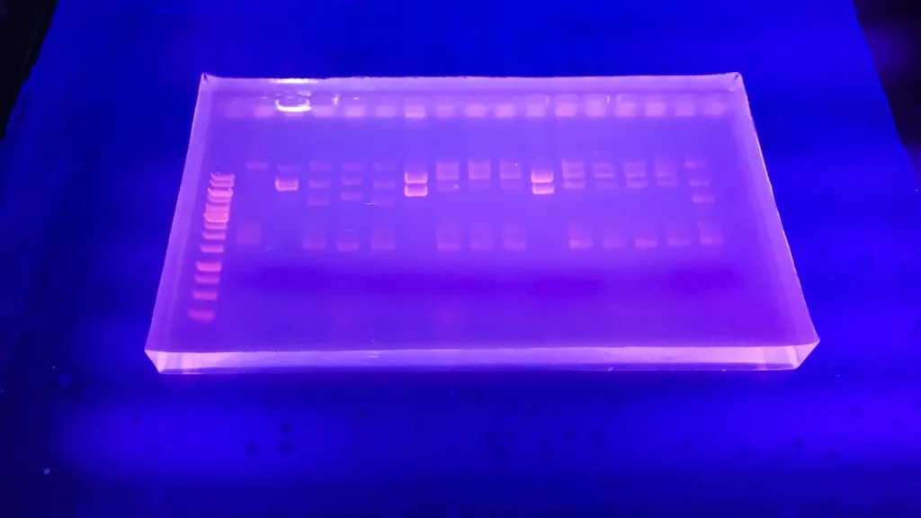
Polymerase Chain Reaction (PCR) is a foundational molecular biology technique that amplifies specific DNA segments exponentially, enabling detection, analysis, and cloning. Invented by Kary Mullis in 1983, it mimics DNA replication in vitro using a thermostable DNA polymerase (e.g., Taq from Thermus aquaticus), primers (short oligonucleotides flanking the target sequence), nucleotides (dNTPs), and a buffer. The process cycles through three steps in a thermal cycler:
- Denaturation (94–98°C): Double-stranded DNA (dsDNA) melts into single strands.
- Annealing (50–65°C): Primers hybridize to complementary sequences on the target strands.
- Extension (72°C): Polymerase synthesizes new strands by adding dNTPs to the primers.
Typically, 25–40 cycles yield billions of copies from trace starting material. Products are visualized via gel electrophoresis, often with ethidium bromide staining, appearing as bands of expected sizes.
In the context of confirming mutations from transposon imprecise excision in Drosophila, PCR is invaluable for detecting deletions. Transposons like P-elements insert into the genome and can be mobilized by transposase, sometimes excising imprecisely—removing flanking genomic DNA and creating deletions (e.g., 100 bp to several kb). To verify:
- Design primers flanking the original insertion site (e.g., one upstream and one downstream of the transposon).
- In wild-type or precise excision flies, PCR yields a large product including the transposon (~2–3 kb for P-elements).
- In imprecise excision mutants with deletions, the product is shorter (reflecting lost DNA) or absent if the deletion removes a primer site.
- Compare band sizes on gels against controls; sequence products for precise breakpoints.
This “deletion-mapping PCR” confirms loss-of-function alleles, aiding gene function studies. It’s cost-effective, rapid, and scalable for screening fly stocks post-excision screens.
Real-time quantitative PCR (qPCR) differs from standard (endpoint) PCR by monitoring amplification in real-time via fluorescence. It uses dyes (e.g., SYBR Green) or probes (e.g., TaqMan) that emit light proportional to dsDNA during each cycle. This quantifies initial template amounts via cycle threshold (Ct) values, enabling absolute or relative gene expression analysis. Unlike standard PCR, which only assesses final products, qPCR provides kinetics data, detects low-copy targets sensitively, and avoids post-PCR handling (reducing contamination). However, it’s more expensive and requires specialized instruments.

Leave a Reply