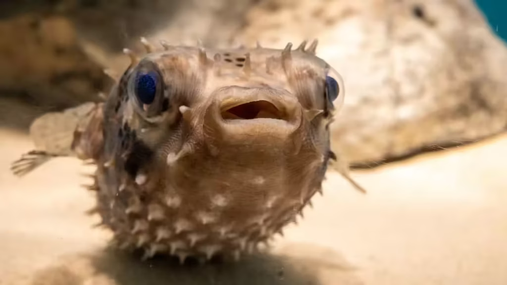
Tetrodotoxin (TTX) is a potent neurotoxin renowned for its role in food poisoning and scientific research. It functions by selectively blocking voltage-gated sodium channels (VGSCs) in nerve and muscle cells. These channels are crucial for generating action potentials—the electrical signals that propagate along neurons and trigger muscle contractions. By binding to the channel’s outer pore, TTX prevents sodium ions from entering the cell during depolarization, halting impulse transmission. This leads to rapid paralysis, respiratory failure, and potentially death in affected organisms, as seen in human cases of pufferfish poisoning. TTX’s high affinity (nanomolar concentrations suffice) and specificity make it a valuable tool in neuroscience for studying ion channel dynamics and synaptic transmission.
TTX originates from symbiotic bacteria, primarily species like Pseudoalteromonas, Vibrio, and Shewanella, which produce the toxin. These microbes inhabit the tissues of various marine animals, conferring the toxin’s presence as a defense mechanism. The most famous source is pufferfish (family Tetraodontidae), where TTX accumulates in organs like the liver, ovaries, and skin—hence the Japanese name “tetrodotoxin” from Tetrodon (pufferfish genus). Other sources include blue-ringed octopuses (Hapalochlaena spp.), certain newts, frogs (e.g., Atelopus toads), and even some starfish and flatworms. The toxin isn’t synthesized by the animals themselves but acquired through diet or bacterial symbiosis, with levels varying by species, geography, and season.
Calcium imaging
Calcium imaging in Drosophila neuroscience uses genetically encoded calcium indicators (e.g., GCaMP variants) to monitor changes in intracellular Ca²⁺ concentration as a proxy for neuronal activity. These sensors fluoresce brighter when bound to Ca²⁺, allowing real-time visualization of neural responses via two-photon or confocal microscopy in live, dissected, or intact brains.
Tetrodotoxin (TTX) is frequently added to the bath solution (typically 1–10 µM, sometimes up to 1 mM with pre-incubation) during ex vivo whole-brain or ventral nerve cord preparations for calcium imaging.
What TTX Does in This Context
TTX blocks voltage-gated sodium channels, preventing action potential generation and propagation. This pharmacologically silences most sodium-dependent spiking activity across the brain network, suppressing spontaneous firing, polysynaptic transmission, and circuit-driven oscillations.
Key Advantages and Applications
- Isolates direct, monosynaptic, or subthreshold responses — In techniques like TERPS (TTX-Engineered Resistance for Probing Synapses), TTX silences the entire network except presynaptic neurons engineered to express TTX-resistant sodium channels (e.g., NaChBac) paired with optogenetic activators. Calcium imaging then detects postsynaptic responses only from direct synaptic inputs, confirming monosynaptic connectivity without confounding network effects.
- Reduces background noise and artifacts — Spontaneous activity, metabolic fluctuations, or network oscillations can obscure stimulus-evoked signals or baseline stability. TTX quiets these (e.g., reducing variance in GCaMP/jRGECO signals), providing cleaner baselines for studying evoked responses, local computations, or direct pharmacological effects.
- Probes activity-independent or presynaptic mechanisms — TTX helps distinguish spike-dependent vs. subthreshold Ca²⁺ dynamics (e.g., in nonspiking interneurons or dendritic compartments), or tests whether observed Ca²⁺ transients rely on network spiking (e.g., in mushroom body Kenyon cells or dopaminergic neurons).
- Facilitates ex vivo functional studies — In dissected brains, TTX stabilizes preparations for long imaging sessions, revealing intrinsic properties like tonic activity contributions or hormone/neurotransmitter effects independent of circuit drive.

Leave a Reply